OPTICAL CO-DESIGN
Light, tuned for the retina.
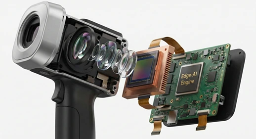
Proposed 530 nm and 590 nm dual-wavelength LED illumination, with a 45° field-of-view optical design. Contrast and imaging performance still need to be tested.
Meet DRADADSS. A proposed handheld fundus camera bringing fully offline Edge AI to diabetic retinopathy screening.
In development · Concept and targets from our pitch deck. Clinical validation and regulatory review remain ahead.
01 / THE CHALLENGE
Screening needs to fit the place where care happens.
The deck identifies a practical gap: retinal screening can depend on specialist access, costly equipment and reliable connectivity. Those dependencies can be difficult to meet in primary care and outreach settings.
02 / THE TECHNOLOGY
DRADADSS brings optical engineering and embedded AI into a single proposed system, designed to work without an external PC or a cloud connection.
OPTICAL CO-DESIGN
Proposed 530 nm and 590 nm dual-wavelength LED illumination, with a 45° field-of-view optical design. Contrast and imaging performance still need to be tested.
EMBEDDED EDGE AI
Planned 8-bit quantized models on custom edge hardware. The engineering goal is inference in under 3 seconds with power draw below 8 W.
LOCAL BY DESIGN
The proposed architecture uses local processing and AES-256 encrypted record storage. Privacy safeguards and security require implementation and verification.
Optical assembly + Edge hardware + On-device models + Local records
03 / THE PROPOSED WORKFLOW
A workflow being designed around frontline health workers. Training needs, usability and clinical performance will be evaluated during development.
A proposed non-mydriatic optical assembly acquires a fundus image in a compact handheld form.
OPTICAL IMAGE ACQUISITIONEmbedded AI is intended to assess the image locally, without sending it to a cloud service.
FULLY OFFLINE DESIGN GOALA planned 5-inch display presents grading and prompts, supporting clinical review and a next-step care decision.
12-LANGUAGE PROMPT TARGETIntended screening grades span no DR, non-proliferative DR severity levels and proliferative DR. These outputs are proposed and have not been established as validated diagnostic results.
04 / ENGINEERING GOALS
Our proposed specifications set the direction for development. They are targets, subject to prototype testing, validation and design changes.
Indicative development goal.
Final pricing and availability are not established.
All values above are proposed in the pitch deck; they are not measured product specifications.
05 / WHERE IT COULD HELP
Three intended care settings guide the product concept. Deployment will depend on validation, permissions and suitable clinical workflows.
PRIMARY HEALTH CENTRES
The intended PHC use case brings retinal image capture and on-device assessment to frontline care, with local records and regional-language prompts as design goals.
CLINICS & CARE NETWORKS
The proposed integrated device could support screening conversations in clinics. Clinical partners would help define safe use, review responsibilities and referral pathways.
COMMUNITY OUTREACH
Handheld weight and battery-life targets are intended to support community screening and mobile outreach, subject to field usability testing and a defined follow-up pathway.
06 / THE DEVELOPMENT ROADMAP
The deck outlines a progression from laboratory bench concept to alpha hardware and a clinical pilot. Milestones below are planned, not reported as completed.
MONTHS 01–03
Characterize the 45° optical assembly and calibrate the dual-wavelength LED system.
ENGINEERINGMONTHS 04–06
Implement quantized models and test inference latency and power draw on a unified PCB.
INTEGRATIONMONTHS 07–09
Assemble handheld units, test thermals and battery life, and carry out IEC 60601 electrical pre-testing.
PROTOTYPE TESTINGMONTHS 10–12
Proposed prospective validation with 300 patients against dilated examinations, and planned CDSCO technical dossier submission.
PLANNED VALIDATIONEvidence before deployment. The supplied deck does not establish completed clinical validation, regulatory approval or certified compliance. A dossier submission is a planned step and does not constitute approval.
07 / THE PEOPLE BEHIND THE VISION
The founder and advisory expertise presented in the pitch deck bring together embedded systems, optical engineering and retinal care.
FOUNDER & CHIEF TECHNOLOGY OFFICER
Experience in robotics, autonomous navigation and embedded edge systems, with prior work in vision-guided surgical robotics and FPGA-accelerated computer vision.
Building at the intersection of optical engineering and on-device intelligence.
TRISHITA RESEARCH LABRetired Professor, IIT Kanpur
Optical system design, image processing and hardware–software integration.
Professor (Retina Vitreous), Department of Ophthalmology, King George’s Medical University, Lucknow
08 / BUILD WITH US
Proposed support for a 12-month development and validation programme.
Help shape the proposed validation study, screening workflow and clinical review process.
Explore optics, embedded electronics, low-power AI and prototype development.
Discuss the development plan and proposed institutional hardware and pay-per-screen model.